
Patient History
Patient is a 59-year-old female who presented with atypical chest pain and dyspnea. Patient’s medication chart included Lopressor, Amlodipine, Captopril, Lasix, Aspirin, Zocor, and Bupropion. Time between SPECT and PET exam was 12 days.
Body Habitus
Height: 5’5”
Weight: 140lbs
BMI: 23.3 Kg/m2
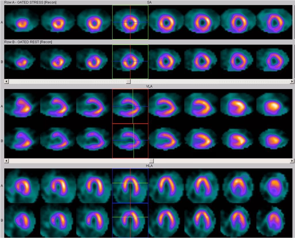
SPECT Images
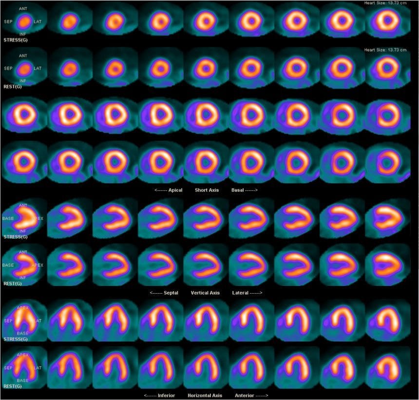
PET Images
| Protocol Characteristics | ||
| Protocol | SPECT | PET |
| Mode of Stress | Adenosine (4 minutes) | Dipyridamole (4 minutes) |
| Clinical Response | Non-ischemic | Non-ischemic |
| Blood Pressure Response | Normal | Normal |
| ECG Response | Negative* | Negative* |
| Radiopharmaceutical | Tc-99m Sestamibi | Rubidium-82 |
| Rest/Stress Dose | 12mCi/30mCi | 36mCi/36mCi |
| Gated | Yes | Yes |
* Patient had normal sinus rhythm and non-specific ST-T wave abnormalities on the resting ECG prior to the MPI studies
Findings
SPECT MPI Report
There was a small defect of moderate intensity in the mid to apical anterior wall that remained fixed on the rest images and most likely is due to breast attenuation artifact; however a non-transmural myocardial scar cannot be excluded.
PET MPI Report
There were no regional perfusion defects seen on the stress or rest images. The patient’s PET/CT test results are normal and suggest no evidence of flow-limiting CAD. The results suggest that the previously described fixed anterior wall defect (her prior SPECT study) is likely to represent an attenuation artifact.
Note: Cardiac catheterization was not performed because the PET MPI study was normal.
Reference: Case study courtesy of Medical Imaging & Technology Alliance (http://www.medicalimaging.org/about-mita/detail-kits/). The report was prepared by Dr. Marcelo DiCarli at Harvard/Brigham & Women’s Hospital in Boston, MA.