
Patient History
Patient is a 77-year-old female who presented with atypical chest pain. Past medical history includes hypertension. ECG shows normal sinus rhythm and nonspecific T-wave abnormalities. Patient’s medication chart included Atenolol, Famotidine, Aspirin, and Lovenox®. Time between SPECT and Cardiac PET exam was 14 days.
Body Habitus
Height: 5’1”
Weight: 160lbs
BMI: 31 Kg/m2
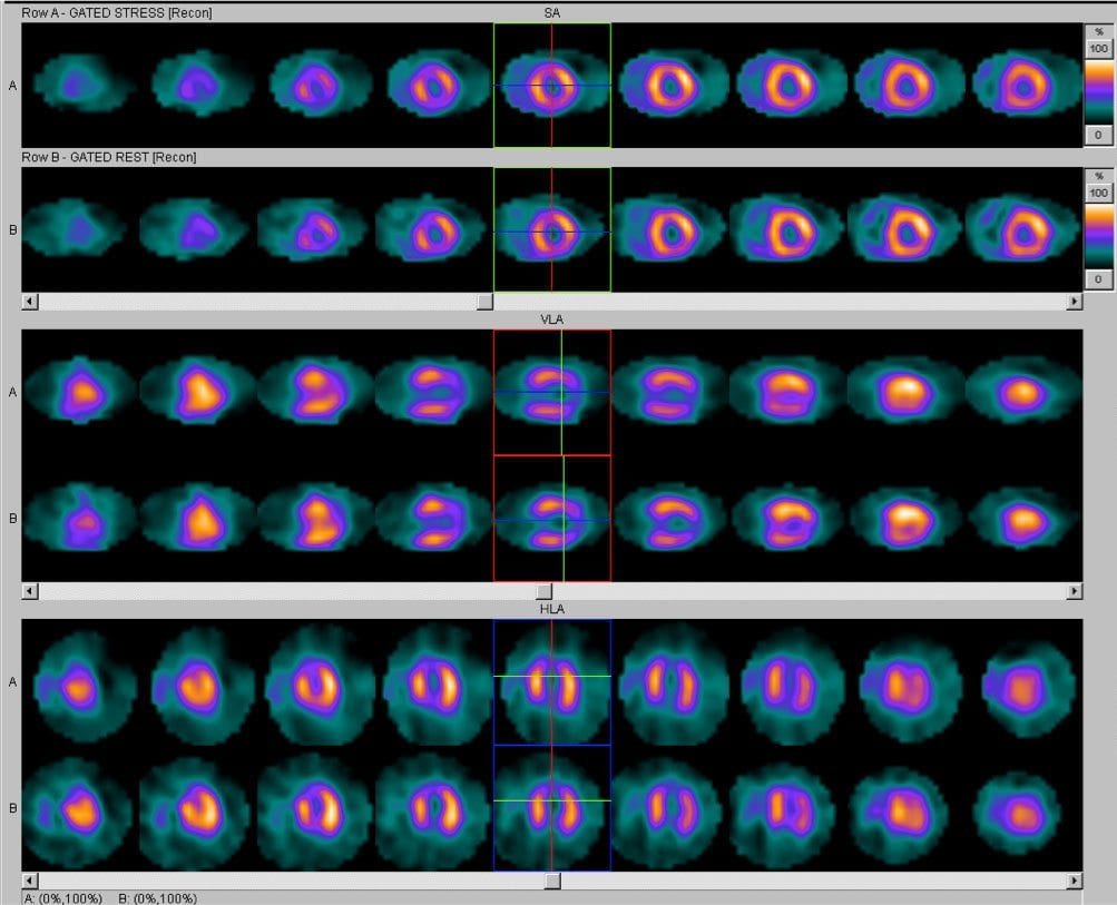
SPECT Images
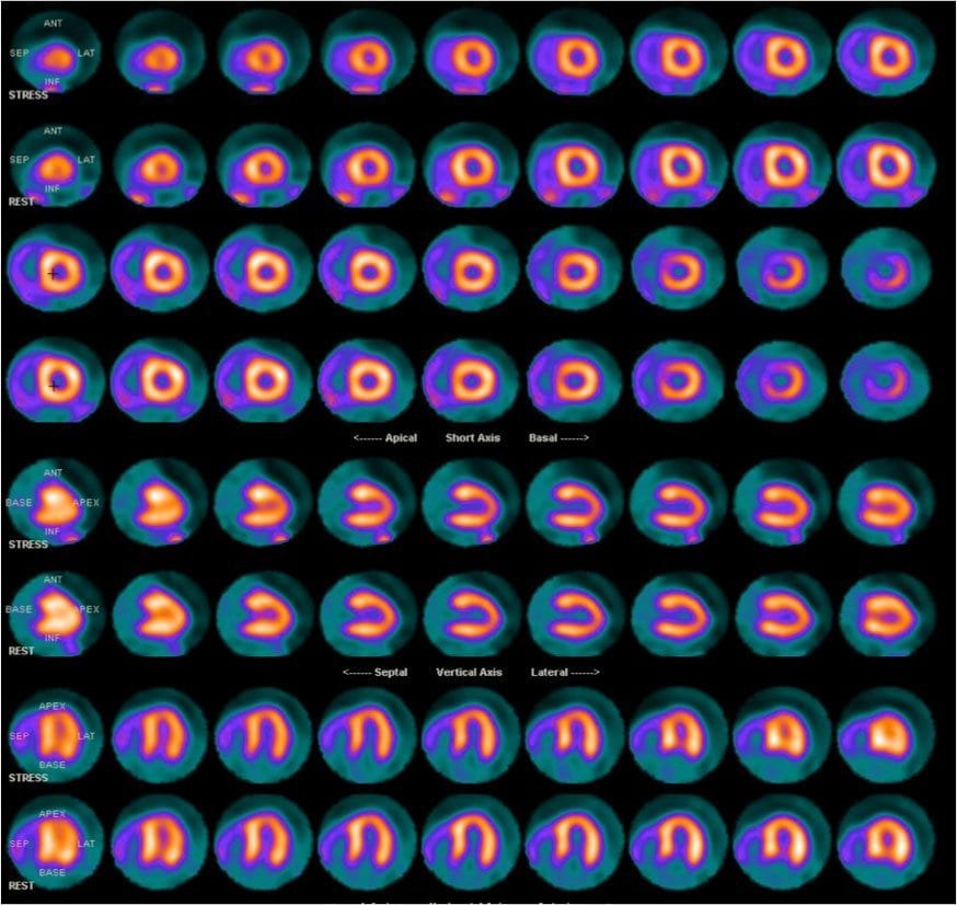
PET Images
| Protocol Characteristics | ||
| Protocol | SPECT | PET |
| Mode of Stress | Adenosine (4 minutes) | Dipyridamole (4 minutes) |
| Clinical Response | Non-ischemic | Non-ischemic |
| Blood Pressure Response | Normal | Normal |
| ECG Response | Negative | Negative |
| Radiopharmaceutical | Tc-99m Sestamibi | Rubidium-82 |
| Rest/Stress Dose | 11mCi/33mCi | 60mCi/60mCi |
| Gated | Yes | Yes |
| Length of Exam Time | 2.5 hours | 40 minutes |
Findings
SPECT MPI Report
Since the SPECT images shown here could not be read with confidence, the patient was subsequently referred for a PET scan.
PET MPI Report
The PET images demonstrated normal myocardial perfusion with interpretive certainty.
Note: Cardiac catheterization was not performed because the PET MPI study was normal.
Reference: Case study courtesy of Medical Imaging & Technology Alliance (http://www.medicalimaging.org/about-mita/detail-kits/). The report was prepared by Dr. Marcelo DiCarli at Harvard/Brigham & Women’s Hospital in Boston, MA.