
 Image Quality
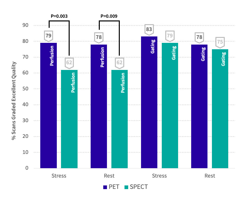
PET Myocardial Perfusion provides excellent spatial resolution and attenuation correction. The enhanced image quality for PET MPI vs. SPECT is due to:
- Higher count rates (240% increase over SPECT)
- Improved spatial resolution
- Routine and robust attenuation correction on all scans
- Images can be sliced at 3mm (PET MPI) instead of 6mm (SPECT) to better detect multi-vessel disease
Diagnostic Accuracy
PET myocardial perfusion has a 95% sensitivity and 95% specificity for detecting coronary artery disease, while SPECT myocardial perfusion has an 87% sensitivity and 73% specificity for detection.
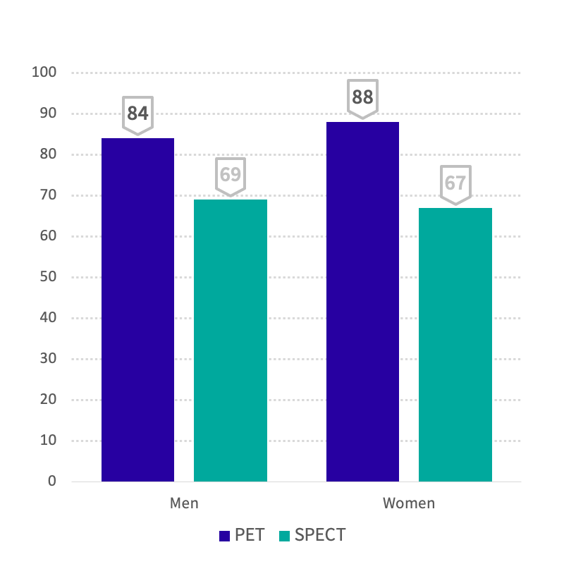
Diagnostic Accuracy by Gender
P=0.55 (MEN); P=0.009 (WOMEN)
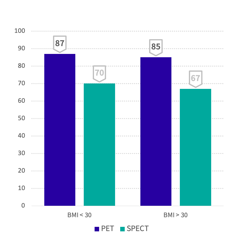
Diagnostic Accuracy by BMI
P=0.05 (BMI<30); P=0.02 (BMI>30)
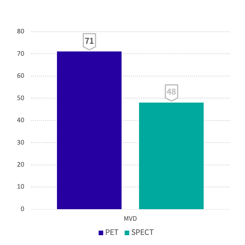
MVD Sensitivity
P=0.03
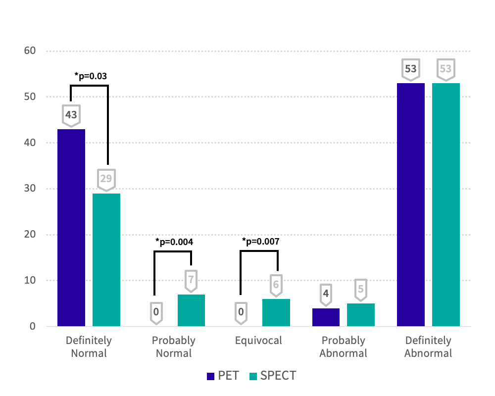
Degrees of Interpretive Certainty
Interpretive Certainty
Better images means greater diagnostic confidence. The literature suggests that PET Myocardial Perfusion images can be read with 96% interpretive certainty vs. 82% with SPECT.
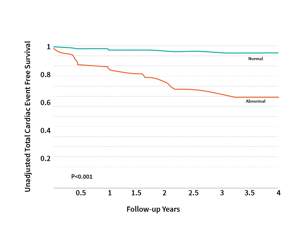
Prognostic Value
Obese patients who are often difficult to image with SPECT had excellent outcomes using PET MPI with Rb-82. The graph below has a curve that shows the difference between survival, free from any cardiac events, for patients with a normal vs. an abnormal PET MPI scan. This study also went on to show that for hard cardiac events (cardiac death & MI), patients with a normal PET scan had a 0% annualized event rate, which is quite impressive.
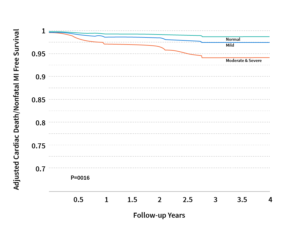
The graph below depicts the prognostic value in all patient types using Rb-82 PET MPI, which is ultimately useful in making patient management decisions.
Rubidium-82
Half life is 75 seconds; dose range is 20-60 mCi. Rb-82 has a high myocardial extraction fraction at peak stress flow; tracer uptake is more proportional to myocardial blood flow and facilitates better detection of disease.
Pharmacokinetics
PET MPI uses higher energy tracers 511keV v. 140KeV for SPECT; this leads to higher count rates and improved image quality.
Artifacts
PET MPI offers attenuation correction on all scans, reduces ambiguity, enhances interpretive certainty
Scan Time Efficiency
30 minutes for PET MPI for gated rest/stress v. 2.5-4 hours for SPECT
PET MPI v. SPECT
| Characteristics of SPECT vs. PET MPI | ||
| PET | SPECT | |
| Availability | Limited | Wide |
| Attenuation correction | Accurate | Less accurate |
| Spatial resolution | 5–7 mm | 12–15 mm |
| Protocol | < 1 hour | 2 days |
| Radiation | < 10 mSv | > 10 mSv |
| Images | Quantitative | Qualitative |
| Hybrid with CT | Yes | Yes |