Patient History
Patient is an 82-year-old male who presented for preoperative cardiac evaluation prior to hip replacement surgery. Past medical history includes hypertension. Patient’s medication chart included ASA and Bisoprolol Fulmate.
Body Habitus
Height: 5’10”
Weight: 210lbs
BMI: 30.1 Kg/m2
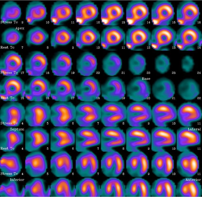
SPECT Images

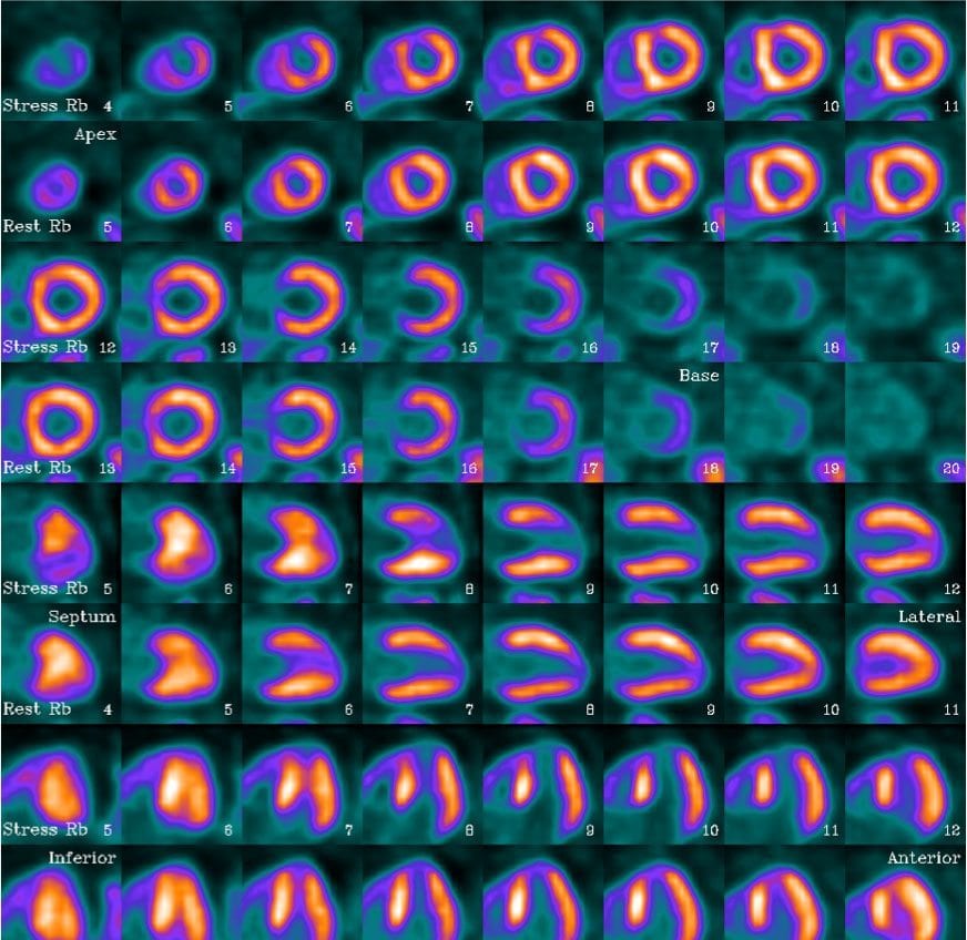
PET Images

| Protocol Characteristics | ||
| Protocol | SPECT | PET |
| Mode of Stress | Dipyridamole | Dipyridamole |
| Clinical Response | Non-ischemic | Non-ischemic |
| Blood Pressure Response | Normal | Normal |
| ECG Response | Negative | Negative |
| Radiopharmaceutical | Tc-99m Sestamibi | Rubidium-82 |
| Rest/Stress Dose | 10mCi/33mCi | 47mCi/47mCi |
| Gated | Yes | Yes |
| Length of Exam Time | 2.5 hours | 40 minutes |
Findings
SPECT MPI Report
Fixed defect is noted at the apex, which does not move or thicken appropriately and likely represents a scar. There is no SPECT evidence of ischemia. LV ejection fraction of 40 percent (normal above 45 percent).
Note: At a BMI of 30, this patient is just over the line for being considered obese and turns out to present a challenge for SPECT during this pharm stress study.
PET MPI Report
The PET images demonstrate the above-described apical scar pattern but also demonstrate a mild to moderate anterior ischemic pattern involving the distal half of the anterior segment. This is suggestive of mild peri-infarct ischemia. The LV ejection fraction in the PET study is 53% at rest rising to 57% with pharmacologic stress (normal left ventricular function). Statistically, the likelihood of a perioperative event is still fairly low.
Note: Even though this patient received pre-op clearance for hip surgery, the detection of ischemia on the PET study provided prognostically useful information to assist in the management of this patient’s progressive CAD.
Reference: Case study courtesy of Medical Imaging & Technology Alliance (http://www.medicalimaging.org/about-mita/detail-kits/). The report was prepared by Dr. Jim O’Donnell at University Hospitals Case Medical Center, Cleveland, OH.
